Orange Lab
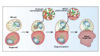
![Diagram showing the critical steps of the NK cell mediated cytotoxicity. During the first stage, NK cell approaches and recognizes a target cell that needs to be eliminated.](https://www.pediatrics.columbia.edu/sites/default/files/styles/cola_media_200_16_9/public/media/images/2018-10/20180924_critical-steps-nkcropped.jpg?itok=EExLOTqx 200w, https://www.pediatrics.columbia.edu/sites/default/files/styles/cola_media_260_16_9/public/media/images/2018-10/20180924_critical-steps-nkcropped.jpg?itok=n6yRGTPS 260w, https://www.pediatrics.columbia.edu/sites/default/files/styles/cola_media_320_16_9/public/media/images/2018-10/20180924_critical-steps-nkcropped.jpg?itok=XaXfMZVJ 320w, https://www.pediatrics.columbia.edu/sites/default/files/styles/cola_media_400_16_9/public/media/images/2018-10/20180924_critical-steps-nkcropped.jpg?itok=s4tU8MdC 400w, https://www.pediatrics.columbia.edu/sites/default/files/styles/cola_media_520_16_9/public/media/images/2018-10/20180924_critical-steps-nkcropped.jpg?itok=9V05vq9G 520w, https://www.pediatrics.columbia.edu/sites/default/files/styles/cola_media_640_16_9/public/media/images/2018-10/20180924_critical-steps-nkcropped.jpg?itok=fM7vB5Rd 640w, https://www.pediatrics.columbia.edu/sites/default/files/styles/cola_media_800_16_9/public/media/images/2018-10/20180924_critical-steps-nkcropped.jpg?itok=VTgeLiPn 800w, https://www.pediatrics.columbia.edu/sites/default/files/styles/cola_media_1040_16_9/public/media/images/2018-10/20180924_critical-steps-nkcropped.jpg?itok=_OK0Y3HT 1040w, https://www.pediatrics.columbia.edu/sites/default/files/styles/cola_media_1280_16_9/public/media/images/2018-10/20180924_critical-steps-nkcropped.jpg?itok=wLRpD0w- 1280w, https://www.pediatrics.columbia.edu/sites/default/files/styles/cola_media_1600_16_9/public/media/images/2018-10/20180924_critical-steps-nkcropped.jpg?itok=Z217e0oo 1600w)
Location and Contact Information
Principal Investigator
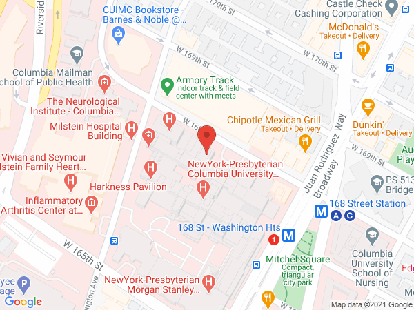
The Orange Lab is located on the tenth floor of the Vagelos College of Physicians and Surgeons (VP&S) Building at Columbia University Irving Medical Center. We share space with the Mace Lab and our groups collaborate closely to dissect mechanisms of human natural killer (NK) cell maturation and function.
Cytotoxic activity of NK cell
In this movie acquired by confocal fluorescence microscopy, a Natural Killer cell was stained with a red fluorescent dye to highlight the lytic granules contained throughout the cytoplasm at the beginning of the movie. Following the recognition of a target cell (stained using a yellow dye), the lytic granules are rapidly converging and polarizing against the interface between the two cells. The content of the granules is transferred onto the target cell and induces the death of the recipient cell. This mechanism is shown by the intake of a purple dye that is infiltrating cells and binds to the DNA of cells that have been compromised. Total duration of the movie 90 minutes. Experiment acquired by Hsiang-Ting Hsu, PhD.